
Thickened edematous wall of the gall bladder ( 7 mm - normal is less than 3 mm )
The patient had chronic ascites
Link: http://www.ultrasound-images.com/gb-wall.htm
Diffuse GB wall thickening is the hallmark of acute cholecystitis. Remember that this is an unspecific finding, and can be caused by a wide spectrum of conditions (see below).
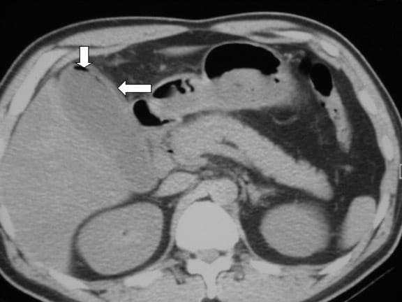
Postprandial state (collapsed GB) can mimic wall thickening. On CT the GB wall appears as a thin rim of contrast enhancing soft tissue.
When is the wall thickened?
Thicker than 3 mm, layered structure.

Acute cholecystitis:
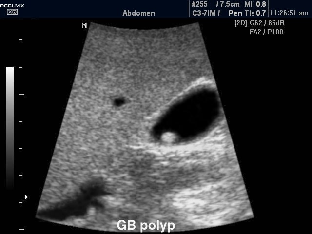
US- GB wall in acute cholecystitis appears as a thin hypoechoic line sandwiched between two echogenic lines.

CT- thick wall with hypodense outer layer (subserosal edema).
§ Try to look for -obstructing calculus, thick sludge
- Hydropic distension (not compressible)
- Murphy's sign (pain)
- Hyperemia in CDS
- Fluid arround the GB
Acalculus cholecystitis:
In critically ill, due to increased viscoscity (sludge) from fasting (parenteral nutrition.

Chronic cholecystitis:
Stones causing only transient obstruction (and symptoms) - leading to little inflammation and fibrosis.
Xanthogranulomatous cholecystitis:
Intramural hypoechoic/hypoattenuating foci of abscess and inflammation.
§ Differentiantion from GB cancer is important in this case!

Porcelain GB:
Rare, due to chronic inflammation, mural calcification.


GB carcinoma:
Wide range of imaging findings (polypoid intraluminal mass, infiltration, diffuse mural thickening, etc..)
§ Difficult to differentiate from xanthogranulomatous cholecystitis.
Adenomyomatosis:
Muscular hypertrophy, intramural polyps. Benign condition, no treatment needed.
Comet tail reverebation artefacts from cholesterol stones are pathognomic.
Emphysematous cholecystitis:
Very ill patient.
In liver cirrhosis:
Diffuse GB wall thickening. Uncertain pathophysiology, but probably due to increased portal pressure.
Drug induced hepatitis:
Can also lead to GB wall thickening.
CHF, pancreatitis (direct spread of infl.), mononucleosis, hepatitis, AIDS can all cause thickening in the GB wall.
What else to check?
Labs (CRP, WBC), fever, onset after heavy meal, liver enzymes ( bilirubin, GOT, GPT, ALP)
Dimensions:
100 x 40 mm less than 3 mm wall thickness
No comments:
Post a Comment