Method of Beatson and Pearson; JBJS 1966;
- AP of Foot:
- taken in 30 deg plantarflexion, with the x-ray tube directed
30 deg from the perpendicular;
- lines are drawn longitudinally thru the talus parallel to its
medial border and thru the calcaneus parallel to its lateral
border;
- on AP view, talocalcaneal angle should be between 25-40 deg;
- angle more than 35 deg indicates valgus;
- angle less than 20 deg indicates varus;
- Lateral of Foot:
- taken with the foot in 30 deg of flexion;
- x-ray beam should be perpendicular to both malleoli;
- lines are drawn longitudinally thru the central axis of talus
and parallel to the lower border of the body of the calcaneus;
- parallelism of the calcaneus and talus is key;
- this is recognized as a decr in lateral talocalcaneal angle
which is normally 30-50 deg;
- this does not increase on max dorsiflexion view;
- on lateral view, this angle should be between 35 - 40 deg;
- Forced dorsiflexion lateral:
- will show an angle smaller than nl (35-50 deg);
- w/ club foot, axes of talus & calcaneus becomes more parallel;
- most reliable roentgenographic view is the lateral projection,
usually with the foot in maximum dorsiflexion.
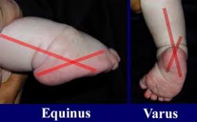
- in clubfoot there is no convergence of talocalcaneal region
(parallel alignment), and the tibiocalcaneal relationship
reveals equinus;
- plantar flexion at the ankle (equinus)
- forefoot and hindfoot inversion (varus);
- Kyte's angles from AP and Lateral views are added together to form
talcalcaneal index;
- in a corrected foot the talocalcaneal index should be greater than 40 degrees;
Talocalcaneal angle
the angle between the long axes of the talus and calcaneus as measured on a weight-bearing radiograph. The talocalcaneal angle on an AP radiograph in a child less than 5 years of age has a normal range 20 - 40. The talocalcaneal angle on a lateral radiograph in a child less than 5 years of age has a normal range 35 - 50. An increased talocalcaneal angle signifies the heel (calcaneus) is in valgus a feature of flatfoot for any cause (flexible flat foot, rigid flat foot or congenital convex pes valgus). Conversely a reduced talocalcaneal angle signifies that the heel (calcaneus) is in varus as in congenital equinovarus, assessment for which is the major role for the talocalcaneal angle.SUMMARY
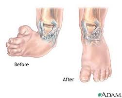
Rocker- bottom foot - Talipes Equinovarus
Vertically oriented talus with increased talocalcaneal angle on lateral view.
Dorsal navicular dislocation at the navicular joint.
Heel equinus.
Rigid deformity.
Frequently associated with : Arthrogryposis multiplex congenita, and spina bifida.
No comments:
Post a Comment